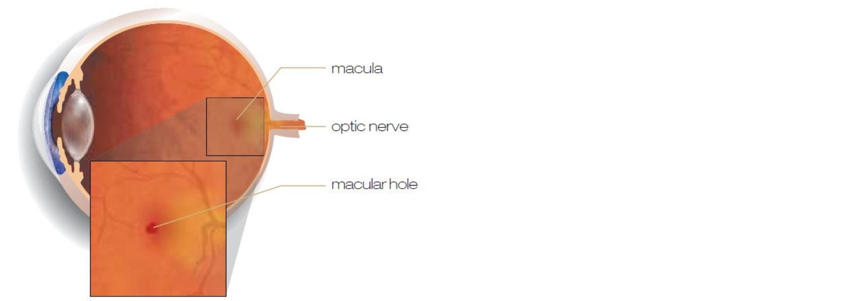
Understanding Macular holes
The retina is a thin layer inside the eye that responds to light and sends visual signals to the brain. The macula is the middle section of the retina. It’s responsible for the central part of your visual field and processes colour and fine detail. When tiny holes or tears appear in the macula, usually because of ageing, your central vision can become distorted, with permanent dark spots in the visual field.

Symptoms and causes
When macular holes first appear, you may notice slight blurring or distortion of vision. As the hole or holes get bigger, they can severely reduce your central field of vision (the area you see when looking straight ahead). You may also notice dark spots or patches in your eye that don’t seem to disappear.
Risk factors
Although there’s no single cause for macular holes, there are factors that may increase your risk.
These include:
- Age – macular holes can happen to anyone but are most common in people in their 60s and 70s.
- Gender – women are more likely to develop macular holes than men.
- Previous macular holes – if you have experienced macular holes in one eye, you are more likely to have them in the other.
- Myopia (near-sightedness) – people with severe myopia are more susceptible.

Diagnosing macular holes
Because the symptoms of macular holes can be similar to other eye disorders, diagnosis involves an imaging test called optical coherence tomography (OCT). Your ophthalmologist gets a clear cross-section of your eye, showing any tears or holes in the macula.
Treatment options
Macular holes don’t always require treatment. If yours are very small and have little effect on your vision, your ophthalmologist may suggest monitoring rather than immediate treatment.
Vitrectomy surgery
If macular holes affect your vision, surgery is an option. The most common surgery is called a vitrectomy. It involves removing the vitreous jelly – the clear, gelatinous liquid that fills the centre of the eye – to access and repair tears in the macula. The surgery is done under local anaesthetic, so you will be awake but won’t feel anything.
During the procedure, vitreous jelly is suctioned out of the eye and replaced with saline solution, air or gas. The surgeon then removes membranes around the macular holes so they can close and heal. A gas bubble is placed in the eye to hold the tear shut and aid healing. After the procedure, the eye will gradually replace the vitreous jelly and return to normal.
Recovery
Recovery from macular hole surgery can take two to four weeks – with at least a week away from work. This is mainly for the gas bubble left in the eye. To keep it in place, you must hold your head in a specific position for some time – usually looking downwards. You must also avoid air travel for at least three weeks after surgery.
In most cases, surgery successfully closes the holes and vision is improved.


