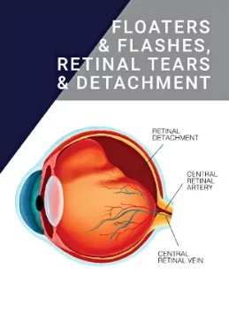
Understanding retinal tears or detachments
Retinal tears or detachments can happen as part of the ageing process or because of trauma or injury to the eye. They can lead to severe vision problems and loss of sight and must be treated quickly.
Risk factors that can lead to retinal detachment
A history of retinal detachment in yourself or a family member
- Aging
- Serious eye injury
- Eye surgery
- Severe myopia (near-sightedness)
- Diabetic retinopathy – a condition that affects the blood vessels in the retina
- Other eye diseases

- A sudden ‘shower’ or rush of floaters (transparent spots or shapes) in the eye
- Flashes of bright light in one or both eyes
- A shadow or dark patch over one part of your vision
If you have any symptoms, seek help from your ophthalmologist or doctor immediately.
Click here to view a video that shows how the retina’s layers can separate, resulting in the retina’s inability to transmit visual signals to the brain.
Click here to view a video that shows how the vitreous shrinks over time, possibly tearing the retina in the process, leading to vision changes. Routine check-ups are advised to avoid severe damage.

Treatment
Retinal tears and detachments can be very serious and must be treated quickly. Prompt treatment may prevent total detachment and the need for surgery.
Plaotocoagulation
This treatment uses a laser to target the tear. The retina reacts to the laser, and the resulting scar tissue seals the tear. It’s usually a safe, painless process.
Cryotherapy
This minor procedure uses extreme cold to seal tears in the retina. A tiny probe is touched to the outside of the eye, and the extreme cold works through to the retina, creating scar tissue that seals the tear. It can be performed under local anaesthesia.
Retinal detachment surgery
If your retina becomes completely detached, there are several surgical options for treatment:
Pneumatic retinopexy
Cryotherapy or laser therapy seals the retina on the wall of the eye to treat straightforward detachments. Then, a gas bubble is injected into the eye to prevent liquid from moving around and hold the retina in place while it heals.
Scleral buckle
Your surgeon will open your eye and reattach the retina before suturing a silicon ‘strap’ to the sclera, or white of the eye. The strap holds the sclera and the retina together, helping the retina stay in place.
Vitrectomy
The most common treatment involves removing the vitreous body and replacing it with gas or oil to reduce pressure on the retina. Laser therapy then reattaches the retina. During recovery, the gas or oil holds the retina in place to heal properly.
Recovery
Retinal surgery has two to four weeks of recovery when your eye may be painful, feel gritty or look red and swollen. You will need someone to drive you home afterwards as your eye will be covered.
If your surgery involves placing a gas bubble in your eye, you will also need to hold your head in a specific position for an extended period to keep the bubble in place. You may need to wear an eye shield at night and use eye drops regularly.
During recovery, avoid:
- Strenuous exercise or heavy lifting
- Air travel if your treatment involved a gas bubble
- Sports
- Dirt, dust or water entering the eye


